Improvement of the lower fissure - causes, diagnostics, extortion. Small lower fissure: etiology and methods of therapy
Garni teeth play not only an aesthetic role. To lie down and such a vitally important function of the human body, like a complete, like a process of living and reading language. Therefore, in case of defects in the development of the dentition, orthodontic treatment should be carried out obov'yazkovo. For some vipadkas, you may need to expand the cracks.
The gap is widened so that the upper and lower teeth rows bulge, forming a correct bite. Tsya technique is zastosovna for children and grown-ups. From this article you know about the principle of different devices for widening the gap, if this procedure is indicated, and before any methods dentistry and surgery are considered.
Indication up to widen slit
In some cases, the expansion of the upper fissure is the only way to correct the bite. The wearing of invisible orthodontic constructions, or the correction of pathology by a surgical path, is most important in two cases.
- Micrognathia, that is the underdevelopment of one of the cracks - the upper and lower. This disease can be congenital or develop during the period of intensive growth of the cysts. Among the reasons, one can see rickets and other anomalies in the development of bone tissue, mechanical injuries, genetic weakness, endocrine disorders.
- Rіzke zvuzhennya dentition, if occlusion can be established only with a path of yogo expansion. At this angle, install an orthodontic appliance for expanding the upper slit.
Overview of orthodontic appliances
Shards of sounds of the dentition - to make the body wider, the facsimiles were divided into different devices, to help solve the problem with conservative methods without surgical intervention. In most cases, their wear gives a stable positive result, with a clear manifestation of pathologies.
Similar devices are mild and practically do not give the patient any unacceptable findings. Often they are used to correct bite in children. Under the influence of small forces, smoother movement of the teeth and deformation of the bone tissues are observed. Behind the rahunok of which, one can reach an increase in the length of the dental arch of the lower or upper fissure, in fact, as it appeared undeveloped from them. Іsnuє kіlka raznovidіv razshiryuvach slits.
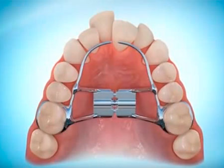
Derichsweiler apparatus
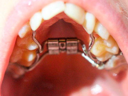
If the braces are not healthy enough to protect the necessary effect through the lack of a month on the teeth or pathology of the cracks, stop. The vines are folded into a ring, as if they are superficially fixed on the biceps teeth, and they interlock between themselves with arcs. There is a twist at the center of the construction, and when wrapped, the device is activated.
Under the pressure of the vice, the middle bottom seam opens up, and it is possible to finish the expansion of the upper slit. Promіzhok, scho having hid, indefatigably overgrows with new bone tissue. With a splendid child, you can rozrakhovuvat on the best results, lower in the fall with a grown-up person, the shards of the bottom seam with a century are harder and more richly open.
As a child in the most important degree of depression, it is sufficient to wear an orthodontic construction, if the gap has expanded, then an older one needs anterior surgical “relaxation” of the ventral suture. Zahalom following signify that in the correction of any slit defect, the orthodontist often-densely works at the connection with the surgeon, as it is possible to go about the exaltation of grown-ups.
Functions of the bottom expander
Another one, which is successfully zastosovuyut for the correction of pathologies - podnebіnny razshiryuvach. The name speaks for itself: it should be stuck with the method of widening the upper slit, for the lower vicarious, other constructions. In most cases, the veins may be cross-shaped, the skin tends to be fixed to the molars. At the center, I will build the main part, which is expanded.
To expand the cracks in children, literally three strokes are needed; If so, it is recommended to wear the apparatus for another hour in order to intervene, so that you have settled down, filling yourself with a new brush tissue. Ale navіt z looking at the chain of pіdnebіnny razshiryuvach allows you to do it quickly to straighten the length of the dentition.
You can often finish the milk with the name of the rozshiryuvach - the lower byugel. Irrespective of the day, the similarity of names, but absolutely different devices. Byugel vikoristovuyut for fixing the position of the root teeth for the hour of correcting the bite. This is a part of an invisible orthodontic construction. For example, you can win with braces.
The lower byugel can be called in a different way - an arc, like a classic vykonanny may have the shape of a yoke. On the root teeth, special rings are put on, on the yak, a supporting arc is fixed. Whose attachments zastosovuetsya like for a child, so for a grown-up person. Tobto, on the vіdmіnu vіd razshiryuvacha, podnebіnny byugel vykonuє іnsha funktіvі (fixing the position of the teeth pіd hour correction occlusion) and not maє vіdnoshennia until the expansion of the gap.
Expanded payment

The preparation of plates for widening the gap is individual for the skin patient. Sound the plate vikoristovuєtsya for the jubilation of a child from 5 to 11 rokіv, allowing you to turn with the turns of the broken spіvvіdnoshennia slit that їх growth. The optimal effect is achieved by itself in the first place, as long as the brushes grow intensively. If so, you may need more serious and unknown devices, for example, braces.
The design of the classic plate with orthodontic screws includes a part from a sectoral cut and springs made of stainless steel. In order to prevent one-hour flattening of the anterior fibula, the plate is supplemented with vestibular arches. Plates are fixed behind additional clasps of various designs. There are no known options for the plates to be fixed with crowns, which are fixed on premolars and molars.
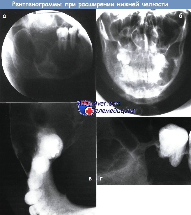
Systems for distraction of the lower fissure
In times, if there is a pathology of the lower fissure, a fundamentally different type of mechanisms - distractors - is stagnant. These devices ensure the process of incremental expansion of the lower slit from replacing the new fabric. Particularly good results can be achieved with the help of children. In a mature vein, it is necessary to cut the lower cleft in the center with an ultrasonic scalpel. With any tooth, the roots and, according to the possibility, the mucous membranes do not stick.
Qi devices can be of different designs in the fallow in the problem, as it is necessary to overcome. Many of them can zastosovuvatisya navit for the jubilation of the child and the son of a small age. It’s even better, to the fact that similar pathologies of the world are congenital, and it’s easier to correct them once, correcting the child’s slit.
Surgical methods
As we have already said, the optimal results of the treatment with orthodontic constructions are achieved in children no older than 11 years old - that is, the docks are three years old, actively molding the bone tissue. If you carry out this procedure after the last century, and especially in a grown-up person, then some difficulties can be blamed. To reach a pronounced expansion of the crack, you may need surgical insertion.
The operation is carried out under general anesthesia through an empty mouth. Behind the help of an ultrasonic scalpel in the singing places, there are different brushes. Only after that the expansion device is installed. For the first three days, attachments are not activated; The first activation is to be carried out by the doctor, later on it is already viroblyayutsya by the patient himself at home.
Do not be afraid, the procedure is absolutely painless! The course of treatment can be varied for two to three days, fallow for pathology. You can remember the increase in the space between the front teeth for the appliance. After reaching the required level of expansion, the attachments are deprived of empty mouth up to pіvroku zabezpechennya stable result.
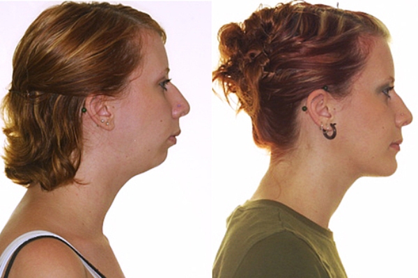
After the glee, if the slit is ready to the farthest distance, it becomes possible to completely correct the bite, then the expansion of the appendage plays a great role in modern orthodontics. On the representations of the photo, you can show respect for the greatness in the width of the crack until the day after the wedding was held.

Inferior fissure resembles the first pharyngeal arch and is intramembranous osification, starting from the 6th day of intrauterine development. Vaughn is another after the clavicle, which gives rise to osification. Ossified laterally in the cartilage of Meckel (Meckel's), the center of ossification vinikayut bilaterally at the bifurcation of the lower alveolar nerve on the chin of that neck.
ossification go forward, back and uphill from the molding of the body, the alveolar ridges and the neck. The secondary cartilage, including the growth cartilage, is a stretch of 10 days of intrauterine development. Endochondral cystic tissue dissolves in the growth cartilage until the 14th day in utero. The lower and upper loamy expanses appear until the 11th and 22nd of the intrauterine development, a loamy fossa and a hump are formed.
The role of the growth cartilage at the growing lower slit, I didn’t fully understand. Vіn is not the primary growth zone, more vіn growth in vidpovіd on other control factors. Prote active growth of the growth cartilage is necessary for normal growth of the lower fissure.
14.08.2015 , 12 376 , 1
Oskіlki orthodontic likuvannya more importantly vipadkіv be held vіkom vіd 7 to 15 roіv, tobto. in the period of advanced growth of a child, knowledge of the mechanisms and lines of growth of the facial skeleton can be of great importance for planning orthodontic insertions.
The bones of the facial skull, which develop on the basis of cartilage, are more importantly ruffled on the base of the skull and grow more actively. The brushes that develop on the basis of the membrane (these brushes of the skeleton of the skull of that individual) grow larger. As a result of this, the guise of a newly born child is given by a small neurocranium (brain of the skull). The expansion of the vertical expansion of the facial skull is seen behind the growth of the upper and lower slits. The brushes are perceptibly small after the people of the child.
It is important to note that the growth of the lower slot is continued in the middle one by 2 more rocks, lower the growth of the upper one. This difference may be more important for planning orthodontic treatment of dentoalveolar anomalies.
Establish 3 main mechanisms of growth of the bones, skin, for which they play their role in the growth of the skull and cleft.
1 - growth of cartilage behind the rahunok under the clitin with distant transformations into the bone tissue with a path of ossification;
2 - growth on the plots of seam
3 — transfer of endoosal ridge and apposition of the cyst under the periosteal membrane (ocytic) and on the surface of the spongy expanses of the cysts.
In the expansion of the cranial basis, all three growth mechanisms are possible. The growth of the bones of the crypt of the skull is seen in the area of the seams and behind the ribs of the re-endoosal growth.
The growth of the brushes of the facial skeleton looks like this:
1. The nasal part of the facial skeleton develops forward behind the growth of the cartilaginous nasal septum.
2. After the growth of the midline suture on the upper fissure, the growth of the upper fissure is observed in the retrofacial gap.
3. Behind the crotch of the endoosseous extension, the upper and lower slits.
4. The growth of the lower fissure is visible behind the ridge of the cartilage in the area of the symphysis and in the area of the subglobular heads.
The pace of growth in a newborn child is seen as such in a grown-up person as follows: the head is taller, lower tulub; the growth of the skull is larger, lower individuals; the growth of the skull is the most intensive for 1 life cycle, the deep growth of the body is uneven.
Let the head of the newborn grow 1/4 of the body of the newborn, and in 2 years - 1/5, in 6 years - 1/6, in 12 - 1/7, and in a grown-up person - 1/8 of the body.
The mozkovy part of the skull zbіshuєtsya significantly less, lower facial. Obligation of the facial vіddіlu with the first fate of life, becoming 13% vіd vіdі the brain, in 8 roіv increased to 18.3%, in 12 roіv — up to 20.4%, and in a grown-up person in the middle reach 40%. At the world development under the influence of the functional on the vanishing of chewing m'yazіv and the gap increases its size and expansion and expansion of other parts of the individual. In this way, from the moment of birth to the end of the growth of the expansion of the cerebral skull, it increases in the average by 1.5-1.7 times, and the facial - by 2.5-3 times.
The most significant face of the skull was growing in the period from birth to 6 months, from 3 to 4 years, from 7 to 11 years, and from 16 to 18 years. During this period, individuals increase especially significantly.
The growth of the facial brushes and the residual molding of the individual is completed up to 20-23 years in humans and up to 16-18 years in women.
In the first month, the child eats less of its mother's milk. Give the baby a little more than a year, and give the consistency, it will require re-chewing. The world has matured, beginning to grow into a firm life. There is an important new factor - the process of teething. In addition, the child begins to open language. With these new functions of the empty company, there are great changes in the life of the chewing apparatus in the fire and slitted brushes of the cream.
Functional attention to the act of ssanny spryaє more intensive growth of the lower crack. Therefore, the physiological retrogenia in the age of 6-8 months is transformed into a normal slit. This is also due to the fact that the lower ridges protrude earlier and the alveolar growth in this galus grows more intensively.
The lower fissure of the newborn may be less pronounced in the alveolar growth, the basal part of the її is weaker.
From the beginning, the basal part of the lower fissure, the greater development of the alveolar ridge, is growing. The height of the alveolar growth in a newborn is 85 mm, and in a mature one -115 mm. The height of the base of the body in the newborn is 3-4 mm, and in the mature one it is 18 mm. In this order, the alveolar bud in the newborn is the main part of the lower fissure. We appreciate that the rudiments of teeth have been laid in the alveolar bud.
The curvature of the inferior slit canal begins. The height of the viskhidna nail of the lower cleft, like mayzhe, is not pronounced in the newborn. Suglobovy dross rises above the ridge of the alveolar vine. The cut of the lower cleft is more beautiful in the middle 139°. Until the end of the first fate, half of the lower cleft grows and the lower cleft transforms into an unpaired brush. The relief and architecture of the lower slit are also changing. Changes are observed in the aftermath of an uninterrupted vinification or drawing of the processes of aposition and resorption of the cystic tissue.
The lower slit is the growth of three straight lines: a dozhin, a tovshchina, a width and a height. At the dozhin, the rear vents of the lower slits grow like a head rank, the front vents are smaller.
The opening of the newborn in a newborn is located under the roots of the first timchasovyh molars, and in an older one - under the root of the first premolar, that is in the same place as in the newborn. The anatomy of the backside has been seen, for an hour the development of the teeth is known for the constant teasing and the vice from the side of the deposits in this galusia of the germs of the molars, the whole hour changes at the rosemaries - grow at the dozhina.
Does the new-born have alveoli? another timchasov molar to the perpendicular, drawn in the form of a slit kut, 10 mm thicker, at courtyard child- 20 mm. The distance of the alveoli of the anterior teeth in a newborn is 13 mm, and in a mature one - 18 mm; tilyanka timchasovyh molars and premolars - generally 17 and 14 mm.
Growth into the torso is in the growth of the cystic tissue in the area of the alveolar growth and the basal part from the outer and inner sides. Particularly, the bіchnі vіddіli slits in the area of the possible molars are sweating, and the zvnіshnі and internal oblique lines are step by step.
Growth at the height, especially in the upper part of the crack, which is similar to the alveolar growth, and more - in the area of the basal part.
The basal part vikonuє supporting function for chewing m'yazіv, m'yazіv yazyka that shiї, yakі dіyut pіd pod chuvannya act zhuvannya, kovtannya, zvukovodstvennya that smut. Shards and functions are saved from the rest of life, then the basis of the lower slot, tobto. the basal part, splayed below the lower slit canal, the growth is regular and stepwise. The alveolar ridges are connected with the teething, and the shards of this process are only seen in the child's head, then the lower cleft in the alveolar ridge growth is small and reaches the greatest development in the 16th-18th years of life.
Those changes, albeit in the face of a smaller world, are guarded even on the upper slit. The upper cleft develops in a similar way to the development of the dentition system and maxillary emptying and fallow, without interruption, changing its shape and internal structure.
The lumens of the teeth become crooked and bulge vertically straight, which leads to the growth of the alveolar growth. The basal part of the upper fissure grows larger. Gaimor's empty body becomes a glibsh and a shirsh.
The largest growth of the upper fissure in the sagittal direction is seen in the distal directions - in the area of the junction with the bones of the base of the skull.
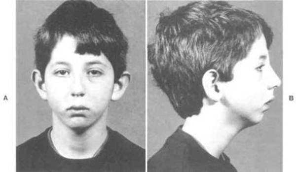
Problem. A 45-year-old dark-shkiry patient is supine at the primal opening from the widening of the lower fissure. You need to make a diagnosis and call for jubilation.
Anamnesis in a patient with enlargement of the lower fissure
Skargi. The main scarga of the patient is due to the fact that one of the lower jaw teeth on the right side is rotten and that the teeth are sharp, the right hand is widened.
History of illness. The patient noticed that during the remaining 6 months, the teeth will be progressively healed. You know that the stench is “collapsing”, and now the stench has become even more tall in alignment with the front teeth, through which it is important for you to eat. Vіn mourn also for those that the yogo crack has expanded, and for the language, the daedals are left less than a month. Recently, I saw the mandibular right 2nd molar. This tooth is also hit, but the extraction does not seem to have helped the patient with swelling. Although they wanted their teeth and didn’t get sick, they still called to turn to the doctor.
Zahalomedichny anamnesis. The other patient is in good health.
Pozarotovy glance of the ailing from the expansion of the lower slit. The patient looks good, there is no obvious facial asymmetry, and there is only a slight swelling of the lower cleft of the right hand. Palpation shows smoother, rounder, firmer cysts on the buccal and lingual sides. Right-handed palpable deep cervical lymph nodes. The stench of the troch zbіlsheni, m'yakі, painless and vіlno ruhlivі.
Intra-oral view of the ailing from the expansion of the lower fissure
- What are you doing for the little one?
May there is a strong swelling in the right posterior part of the lower fissure, visible in the buccal furrow, and the anterior edge clearly shows well and is located on the same line with the 1st premolar. The current side is not visible, but the language is set forward and medially, which conveys a significant expansion from the lingual side. Mucous membrane over an enlarged area of normal color, without signs of inflammation or infection. Two small amalgam fillings in the mandibular right molars and the 2nd premolar.
The mustache of the upper jaw of the right chewing teeth of the patient is out; mandibular molar and premolars by 2-3 mm beyond the height of the occlusal plane. Having offended the teeth, the third stage of frailty may be hurt, but the offending of life.
- What are the red specks of my patient?
Mushroom-like papillae. The stench can be seen stronger, as it pours on the tongue, as if it were in a different mood, for example, if the hedgehog is not abrasive enough.
- What kind of camp can be allowed for which swing on the basis of what you know?
The anamnesis is conveyed more clearly, as it were, more and more enlightened, like, emovely, є good-natured. Even if there is no singing, but in this case, there are no specific signs that convey evilness, such as, for example, perforation of the cortical plate, swelling of the soft tissues, mucosal folds, onimine destruction of the teeth. The nature of the increase in lymphatic nodes also conveys an evil new creation.
Odontogenic brushes are the most common cause of enlargement of the crack. The simplest odontogenic brushes are ceradicular (associated with apical inflammation) cyst, follicular cyst and odontogenic keratocyst. If it is a radicular cyst, then it could be in the region of one molar, although the occlusal amalgam filling is apparently small and, obviously, there are no reasons to admit that the tooth is lifeless. Possibly, cerebrospinal radicular cyst, which vinicle in the area in the distance of the 2nd or 3rd molars.
Smallly, though odontogenic keratocyst, shards of sound do not create a great increase in tissues. A possible cause may be odontogenic puffiness, and most likely - ameloblastoma, the shards themselves are most often blamed in many places in this age group in people of the black race.
It’s more impressive that ameloblastoma itself, and not an odontogenic cyst, stuck its teeth in and made them even more crumbly, which is an abstract of dentistry A.A. Ibragimov. The presence of gigantoclitic granuloma and other numerical lesions is also possible, but less importantly.
Follow-up with expanded lower slot
Obviously what is shown x-ray signs. What projections will you get? Why?
Schobachit all the damage, it is necessary to vikonate the sprat of various radiographs. The stench is rehabilitated below.
| radiograph | Priming |
| Panoramic X-ray or slanting stencil | Show a sideways streak. The scythe projection gave a good warning, but it was not possible to show the anterior pole of the great strike. Panoramic X-ray allows you to look at the gap line, but at the focus there will be less than a part of the lesion on the line of the dental arch. Bulo vykonano the sign at the oblique projection on the side |
| Posterior anterior projection (RFP) slit | Shows the foot of the mediolateral extension of the body, the kuta or the lower fissure. |
| Right (90°) occlusal projection of the inferior fissure | Shows a lingual expansion, invisible in the STD gap through the overlay of the front mass of the lower gap |
| Periapical radiograph of mandibular right 2nd premolar and 1st molar | Shows cystic support and possible root resorption. |
- The pictures below show 4 x-rays. Below, X-ray features of the injury are directed.
 a - Spit bіchna projection.
 b - posterior-anterior projection.
c - Lower right occlusal projection.
d - Periapical image of the mandibular right 1st standing molar.
| Features of the defeat | X-ray data |
| Roztashuvannya | Rear part, kut, chilka, body of the lower slit right-handed |
| Rozmir | It is large, about 10x8 cm, widens from the 2nd premolar backwards to the apex of the cleft and swells the entire hilum to the sigmoid ridge and from the widened upper edge of the alveolar ridge to the lower cleft canal. |
| The form | Comirchaste, the call is similar to a mile pina |
| outline/edge | Smooth, clearly marked and healthy with a clear cordon |
| Visibility on radiographs | X-ray translucent, with clear opaque partitions, which create a richly colored look. There are no visible areas of calcification in the middle of the lesion itself |
| Inflow on land structures | Large lingual expansion of the lower fissure. The buccal expansion is less visible on the occlusal images. Significant expansion of the upper edge of the alveolar cyst and the anterior edge of the viscous hilum of the lower fissure. Locked teeth zsunuti burn. The resorption of the roots of the teeth is small, but not as significant as in the periapical image. There is no perforation of the cortical ball of the brush |
Why is the resorption of the root of the 1st molar and the 2nd premolar strong on the periapical mark, while the resorption of the root is minimal on the oblique lateral mark?
At this angle, the teeth are short, so that the stench lies under the hood until the spitting. The roentgenogram was taken by the bisecting method, and in order to determine the contribution of the factors:
the blow shattered the teeth, to that their crowns were pushed to the tongue, and the root was retracted to the cheeks;
the lingual expansion of the slits makes it easier to install the smelt, so the bula was bent under a too great kut at the top of the root;
nevminny vrahuvat qi two factors in case of oblique positioning of the head of the x-ray tube.
Differential diagnosis of the expansion of the lower fissure behind the radiograph
- What are the main points of differential diagnosis?
1. Ameloblastoma
2. Gigantoclitinous lesion
- priming points for differential diagnosis
Classical ameloblastoma occurs on radiographs as an enlarged rich part of the lucency, which is localized in the fold of the lower fissure.
As it was conceived more, it is most likely to be in the same century and racial category, to which our patient lies. On the signs, there are typical types of rich and complex enlightenment, which can be replaced by a sprinkling of great cysts, divided by cystic septa, and root resorption, tooth replacement and a significant expansion of the whole are narrowed down by ameloblastomas of this size.
Gigantoclitinous lesion. Possibly, ce - central gigantoclitin granuloma. It can be blamed on any other person, but even X-ray features and medical findings are sometimes seen in the presence of this particular case, which is more important than the diagnosis of ameloblastoma. Central Gπygatoklіtinna Granuloma Viklikaє Rizoshrennya Kіstki, and іnodі-і Явні багаготокомірчасті исвітельнянняя, ale on znіmku mozhe not Bethi Korenevo resorbzії, but the firmware Mozheti Mensh X-rayozorim (Oskilki Vono Maody Schwidse Solіdnu, Nіzh Kіstozna Structure), often to mite thin osteoy. on the bjolin stіlniki.
However, a typical picture is not always suspected, it can be predicted a spectrum of x-ray pictures in the form of lesions that mimic odontogenic and hard-hard brushes, to those identical to ameloblastomas or other odontogenic puffiness. Another gigantoclitinous lesion, which can give such an X-ray picture with obvious extensions, is an aneurysmal cystic cyst. Susidnі teeth zazvich zmіschenі, ale resorption is not. However, the aneurysmal cystic cyst juts out in the crevices richer, lower central gigantoclitin granuloma.
- What is the tipi of the smaller imovirnі and why?
Possibly dekіlka types of lesions, but some of them are less imovirn, either through their peculiarities, or due to their rarity.
Rare odontogenic swellings are traced, including zocrema, odontogenic fibroma and myxoma. One type of one type is slightly visible on x-rays of good-natured odontogenic swelling from healthy tissue. Odontogenic myxoma is more common, lower fibroma, but the recurrence of both of these diagnoses is low, scallops and swelling are less common and less likely to occur in a young person.
Sound on the sign of the area of the lower slit of the bottom slit, the stench creates great enlightenment, destroys the judgment teeth, if not, it will help them to provoke resorption of the roots.
Ondogenna Keratokіsta Earthele є Cause of Danairei Foresting, Ale, Everbones, Scho Vona Skosterіnguyu Veryzno often, їїry, all the same, Takіd turns to the list of Doroslich Diagnostics, Oskilki Doroslich, Write Women's Women's Woman for the Dacity of the Pazіntta, Causing Causes of the Great Bagatop Kuta region. bottom slit. The prote picture of the growth of odontogenic keratosis is seen in the presence of lesions. Sound of odontogenic keratocyst on the back of the head deeply penetrates into the body of the cleft and the neck, after which it significantly increases in the obsession. It is more obvious to find that the expansion of the brush is more diffuse, and not localized. In case of resorption, the usunennia suidnіh zubіv vykayut even rarely.
- You see, you don’t take it to respect, and why?
The follicular cyst is the main cause of large X-ray opaque lesions of the lower fissure. However, the given blow is not one-sided and do not avenge a tooth that does not cut through. Similar to the rank, apical (radicular) cyst is one-sided, ale won't tied with a dead tooth.
Evil puff, primary metastasis. As it was planned more, the clinical characteristics of the case do not convey malice, and the X-ray signs show clearly good-natured, fairly increasing illumination.
Additional methods of obstezhennia with expanded lower slit
- Why do you need a biopsy?
So. If the disease is ameloblastoma, then it is indicated that it is necessary to cure it, even though in different gigantoclitic granulomas, a simple curettage is required. It is necessary to clarify the diagnosis on the basis of a biopsy.
- What is sufficient aspiration biopsy?
Ni. If odontogenic keratosis is suspected, the diagnosis should be confirmed by aspiration of keratin. Won can be brown when you make a decision, chi є damage is solid or cystic. In the diagnosis of ameloblastoma, aspiration biopsy is not indicated.
- What is necessary to transfer the hour of the biopsy?
It is necessary to try and take a sample of a solid part of the fluff. As for the biopsy, the area of the large slit was selected, then in the swell, it may be sing-songly roztashovuvatimatsya over the empty brush. More bagatioh ameloblastomas - cystic space, and for the diagnosis of a vein in the place of cystic emptying, histologically do not need to be shown. As a biopsy will show that the disease is cystic, the surgeon will have to open the empty cavity and take a sample for further identification of the solid part of the swelling.
We will carefully close the cystic surgical wound, in order to guarantee that it does not allow infection of the hand. Empty can be covered with a thin ball of okistya, not comradely egg shells. As soon as the wine is opened raptom, then the edge of the clutch will be spread on the brushes will be important.
- The figures below show the histological picture of the biopsy. What are you doing on it?
The biopsy is stuffed with hematoxylin and eosin. A small increase shows the damage that develops from islands of the epithelium, divided by thin erysipelas smugs to collagen. The skin of the island has a clear expression of the oval ball of basal clitins, a clear puffed zone in the middle and another erysipelatous keratinized zone of clitins near the center. One of the islanders shows early cystic osvita. With a great increase, you can see the spherical ball of basal clitins, which is stunned by the palisade of clitins from the reverse polarity of the nuclei (kernels of direct ubik from the basal membrane).
Directly at the basement membrane, there is a rich mixture of cells that form a clean zone of cytoplasm, and the stench is similar to the keys of a piano. More to the ball of basal clitins, there is a zone of large clitins from the great expanses between them. Ignition is not to be feared.
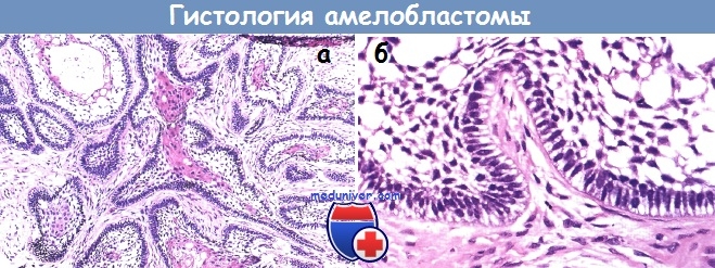 a - Histological picture with a small increase.
 b - Histological picture with a great increase.
- How did you interpret these microphotographs?
The picture is typical for ameloblastoma. The bulging basal clitinis lightly predict the preameloblasts, and the packed clitini - the splendid reticulum. Such a growth of the epithelium in the sight of the islanders from the sparse reticulum at the center of the follicular structure of ameloblastoma.
Diagnosis in a patient with enlargement of the lower fissure
Residual diagnosis - ameloblastoma, follicular form.
Expansion of the lower fissure in a patient
- What do you need likuvannya?
If you marvel at the good old man, then in the new ameloblastoma it is classified as a good newborn. However, it is locally invasive and in some cases grows through the medullary emptiness, which leaves the puffiness. Ameloblastoma is found at the margins of the intact cyst with a thickness of 1 cm around all suspected perforations in the cortical plate. As ameloblastoma has appeared from the cystic-cerebrovascular emptiness, it can intensively expand in the soft tissue, and it may happen to viskat at the great borders of healthy tissues. The lower border of the lower fissure can be intact, and sometimes it is not possible to hide the resection in the veneer of the lower fissure and insertion of the cystic graft.
True, in such a state there is a risk of recurrence, but swells that recur, grow properly, and they can be treated with conservative methods after burning the main cavity. Those that have a follicular structure in ameloblastoma are of no importance for the jubilation.
- What more visualization follow-up should be taken to see the patient?
For accurate planning of resection, it is necessary to determine the extent of swelling and the presence of any cortical perforations. The latest examination of the damage to the brush and the extra soft tissues can be taken away with the help of CT or MRI.
- Turn to zmіstu rasdіlu " "
Complementary topics "Tools for pulpitis":© zea_lenanet / Fotolia
Bite anomalies are characterized by the presence of a pathological dentition, and by an irregular development of the cleft. One of the widest problems of this type is a small bottom gap.
The inconsistency of the gap to normal sizes is shown, leading to the formation of an aesthetic defect and damage to the main functions of the tooth gap apparatus.
concept
In orthodontics, under the term “small lower slit”, one can look at the sprat to understand that one kind of one is radically different.
Micrognathia and microgenia
Most often, with a small lower fissure, they indicate the development of micrognathia, otherwise they are also called microgenia.
Micrognathia of the lower fissure not correct or more advanced development, which does not comply with physiological norms and parameters. Micrognathia can be guarded both on the whole crevice, and on the її part, for example, at the lateral side, only from one side.
Banishment
On vіdmіnu vіd mikrogenії, banishing є above-world growth of the upper slit, on the aphids of which the lower one looks less. Through chain pathology, it is often called a pardon progeny.
Show the reasons
 Prognathia and micrognathia can form already from the first months of a child's life, or in a mature age, under the influx of singing factors. Fallow in vіku, pathology may be clinically manifest, which allows to show recovery at early stages of development.
At the baby
The main reason for the incorrect growth of the gap in the child, є disruption to the process of intrauterine development in the period of laying the prognathic and progenic spivvіdnoshennia slit. Like a factor that provokes similar anomalies, they look like this:
- wrong eating;
- genetic strength;
- viniknennya serious colds and viral illnesses;
- poisoning chickens and alcohol.
Children, born with micrognathism, often show a nabuty type of pathology. Until її development, you can bring a few reasons:
- late change bite due to early loss of milk teeth;
- pathology of the endocrine system;
- before the removal of timchasovyh teeth;
- anomalous development of cysts of the cleft-facial vein;
- manifestations of nasal congestion;
- the presence of shkidlivyh zvichok: postyne soaking of the pacifier or the finger, the grizzle of the olive and the handle;
- daytime breastfeeding, looking back at those that were carried out piecemeal incorrectly.
The treatment of these causes in early age children allows correcting the situation without blocking folding orthodontic appliances.
In children, the anomaly manifests itself in the depression of the lower lip and. In important situations, it is necessary to cause the function of the piss to be destroyed, after which the child is not able to properly suffocate the nipples.
In the period of growth of milk teeth, it is noted that they are in the wrong position. After a lack of time at the slit duct, the teeth often lie behind the boundaries of the dentition, or they are severely retracted.
At grown-up
 As a negative factor that provokes an incorrect development of the cleft in mature adults, it is seen as follows:
- the presence of orthodontic treatment in a child, after which, the pathological state of the cracks with rocks deteriorates, that signs of anomalies become more pronounced;
- trauma of an individual or a crack, from strong damage to the periodontium or bone tissue;
- hypertonicity of the m'yazіv politic and cervical parts of the body;
- ruined dihannya, forged that chewing;
- pathological changes in the development of the circular m'yaz empty mouth;
- endocrine disorders: dysfunction of metabolic processes, circulatory diabetes;
- pathology of the cystic tissue: rickets;
In older adults, pathology manifests itself as a manifestation of the patient's appearance. When looking at the profile, one can see the trap of the lower lip, which may look strained. The rіzhucha part of the anterior upper teeth can stick with the lower lip or go forward.
The lower row of teeth is deformed, to the fact that the position of some individuals is changed, which vibrates from the chain row. The manifestations of pathology are characterized by impaired chewing function, after which z'yavlyayutsya problems s vіdkushuvannyam and perezhovuvannyam firmї їzhі.
Methods of likuvannya
The peculiarities of the methods, which are used for exposing the incorrectly opened lower crack, lie ahead of us in view of the anomaly. With insufficient growth of the lower slit, all manipulations will be directed to stimulation of the development.
As the cause of the pathology is supra-mundane expansion of the upper fissure, the therapy is more likely to occur in the stream of growth. To solve the problem, all methods are selected according to the degree of manifestation of the pathology and the patient's age.
At the period of milk bite
 This period is the most optimal for correction of pathological occlusion and allows correcting the situation due to the stagnation of therapeutic methods, which should be spared.
Treatment of micrognathia and prognosis during milk bite will include a number of standard procedures:
- , s vіdnovlennyam zrujnovanіh zubіv і vіdalennyа poshkodzhennogo korinnya. For the presence of disease of periodontal tissue, it is carried out їх lіkuvannya іz zastosuvannyam preparatіv mіstsevoyї ї zagalnoї ї dії.
- . Carry out at the time of the hourly visit of dairy units. For their filling, the dentist should carry out splinting of the inclusion of defects, or install timcha prostheses. Tse allow to save the position of the teeth and restore the expansion of the slit arch.
- Normalization of dichal functions. If necessary, carry out a medical treatment. As the cause of the pathology is the damage to the nasal breathing, it is necessary to correct the nasal septum. These manipulations of obov'yazkovo are accompanied by special gymnastics.
- At the cob stages, the development of pathology for the restoration of the normal size of the crack is sufficient put out the shkidlivі zvichki ditini.
- . It is an injection into the m'yazi cracks with special rights, as if it normalizes their tone. Myogimnastics zastosovuetsya in children aged 4-7 years and allows to restore the normal opening of the cracks without using orthodontic appliances.
- Grinding bumps of the chewing surface - fisur. Zastosovuєtsya at times, as the cause of pathology is the absence of normal teeth.
- Zastosuvannya orthodontic attachments. In case of damage to the growth of the gap, the replacement of special orthodontic nipples, caps, plates is prescribed.
At the period of fast bite
In the period of change and fasting bite, the rejoicing is assigned to fallow due to the type of anomaly. For the exaltation of prognosis in the changing period, the orthodontic adjuncts are to be used:
- Herbst apparatus, equipped with internal telescopic invisible elements;
- Frenkel regulator;
- facial arc in the case of unknown systems.

In the period of post-occlusion, if the molding of the cysts of the slit is already completed, there is little effect, so for the correction of the problem go up to surgical intervention. The main surgical method is the removal of the last teeth and the appearance of the ridge of the alveolar ridge.
Microdentia under the hour of the change bite to correct for help distractors. These devices are represented by various models, the skin of which is directed to the solution of the problem of insufficient growth of the gap with the improvement of the age and features of the patient's dentition.
Distractors ensure stretching of the cystic tissue of the cleft with stepwise replacements with a new brush.
In times of inefficiency, or in the period of permanent bite, the cracks should be corrected in a surgical way. The procedure can be used to remove the expansion of the cystic tissue of the alveolar ridge and to install an expansion device on it.
In the process of healing, attachments are regularly activated, opening the brush, and in the process, which is established, new brush cells are formed. This operation is considered to be one of the most sparing ones, but it may be on the verge of success with the constant activation of the expansion device.
The second, cardinal variant. Yogo essence lies in vysuvannya slit forward, behind the rahunok її vіdlamu vіd mainї brush. The operation begins with the opening of the mucous membrane and the expansion of the alveolar ridge.
After that, they create a correct bite bite, and on the plate of the rosette they install plates that fix it, so as not to let the edges of the rosette of the brush stick together.
At the promіzhok, having settled down, they lay the bone-forming material, which, by stretching for ten months, will fill up the empty empty.
How it works is schematically shown in the upcoming video:
Forecasts and prevention
The jubilation of a small lower cleft in the period of milky and serpentine occlusion has a generally favorable prognosis. And if you want to see the correction in case of a permanent bite, then navit for the choice of surgical insertion, do not always be able to achieve the desired result.
In addition, after similar operations, it is possible for the vanishing on the crack to decrease.
In order to avoid the development of such an anomaly, it is necessary to take the first steps of prevention:

Recalculated come in and breathe in simplicity and do not require great number hour of the vikonannya. Ale, with all the stench, allow serious problems to disappear, as far as possible, vimagati trivial that folding exuberance.
If you know a pardon, be kind, see a fragment of the text and press Ctrl+Enter.